
Cognitive Neuroscience
- Kaloyan Milanov

- Sep 24, 2020
- 6 min read
Cognitive neuroscience is an area of research focused on determining brain activity with its correspondent actions through neuroimaging technology. This can be used to form localisations of cognitive functions.

The association cortices (located in the parietal, temporal and frontal lobes) form the surface of the majority of the human brain. These cortices are used for complex processing of input, which leads to an output of a specific behaviour. They are also responsible for cognition- the ability to process external stimuli, identify the importance of the stimuli and generate corresponding responses.
The cortex (superficial layer) which covers the cerebral hemispheres is the neocortex, which is formed from six cellular laminae. Each layer is made from a specific type of cell based on the function that needs to be completed. Histologically, they are classified as cytoarchitectonic areas. While each of these areas has specific characteristics, they share some common features:
Each layer has a primary source of input and output
Each layer has connections in the vertical and horizontal axis
Cells with similar functions are radially aligned and are located in all cortical areas
Interneurons form local axons which move horizontally into the cortex and link functionally similar cells.

When a lesion in the temporal lobe is present, it will increase the difficulty of recognition and identification of a stimuli, which suggests that damage to either of the temporal lobes will hinder reorganization, identification and categorization of stimuli. The collective naming of this disorder is agnosias, which has a lexical aspect (matching of wrong cognitive and verbal stimuli) as well as a mnemonic aspect (inability to recall stimuli when exposed successively). Lesions to the right temporal cortex leads to prosopagnosia (agnosia of the faces) and lesions to the left temporal cortex leads to difficulty with language related material.
The prefrontal lobe is the largest lobe in the human brain, with the largest number of cytoarchitectonic areas. This corresponds to the broad functional responsibilities of the lobe.
Language
The frontal and temporal association cortices on the left cerebral hemisphere are used for human language. This form of lateralisation allows specific areas of the brain to specialise in the processing of information for the highest efficiency. The areas of the brain which are cortical representations of language processing are different to the motor areas used to control the muscles used in speaking (larynx, pharynx, mouth, and tongue). While the areas used for processing of visual (written) words, and auditory stimuli are different, they are related to each other.
The ventroposterior region of the frontal lobe (Broca’s area) is used for the production of meaningful words, but not the ability to move the muscles in the mouth and produce words. This area does not control the ability to comprehend language, however, it prevents the ability to organize and control linguistic information. In Broca’s aphasia the individual is not able to express themselves correctly due to the inability to follow the organization of language.
Wernicke’s area is related to comprehension of language. This area can be affected by sensory aphasia, which occurs when there is damage to the auditory association cortices in the posterior frontal lobe. This form of aphasia prevents the linking of objects and ideas to words, but it allows for the formation of fluent speech which is well structured but has no meaning.
Sleep
Sleep is one of the most important physiological processes in the body, as it allows for brain development in the early years (until the age of 20), memory consolidation, and linking of information. The importance of sleep can be seen with a genetic disease known as fatal familial insomnia, which prevents this physiological process from occurring, and leads to impaired memory, cognitive abilities, hallucinations and eventually death.
The circadian rhythm is a periodi, internally regulated process which controls the sleep and wake cycle about every 24 hours. A circadian rhythm is endogenous, which means that it will persist even when the external stimuli (removal of light) remain for long time periods. The rhythms are entrainable, meaning they can be reset by exposure to external stimuli. The circadian rhythm also has temperature compensation, which means that even when kinetic and molecular processes change, the rhythm persists in the individual.
The retinal ganglion cells, which contain melanopsin, have a photoreceptor capability, which sends neural signals to non-image forming areas of the brain such as the suprachiasmatic nucleus. From there, these signals are transmitted to the hypothalamus. When light in the wavelengths of 460nm-480nm is present, the SCN will secrete gamma amino butyric acid, which inhibits neurons that signal the paraventricular nucleus (PVN) of the hypothalamus. Hence, the signal from the pineal gland is inhibited and melatonin is not secreted. When there is no light, the SCN secretes glutamate which allows for PVN transmission of the signal to the pineal gland . The PVN communicates with the superior cervical ganglion in the higher thoracic segments of the spinal cord, hence signalling the pineal gland through sympathetic postsynaptic fibers by releasing norepinephrine. This triggers the pinealocytes to produce melatonin through the activation of the enzyme arylalkylamine N acetyltransferase (AA-NAT).
Melatonin (N acetyl 5 hydroxytryptamine) is synthesized in the pinealocytes when there is a higher concentration of AA-NAT to allow for the conversion of serotonin to intermediates and then to melatonin. The rhythm of melatonin production is internally generated by the the bilateral SCN, where melatonin production is highest in the early morning hours (02:00-04:00), which is then degraded by the liver (converted to 6 hydroxymelatonin by CYP1A2). Melatonin production will also stimulate a decrease in the core body temperature to allow for faster sleep, and receptors in the kidneys for melatonin suggest that urine production is inhibited during the night.
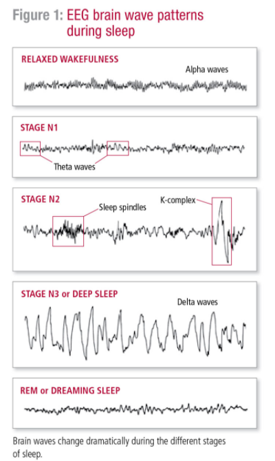
Sleep is composed of two stages - NREM (non rapid eye movement) and REM (rapid eye movement). The NREM stage occurs first and is separated into four different stages. The first stage is associated with alpha and theta waves with a high frequency and low amplitude. As an individual progresses through the successive stages of NREM sleep, the brain waves generated decrease in frequency and increase in amplitude. The first part of Stage I produces alpha waves, which are of low frequency and high amplitude. As an individual progresses through Stage I, there is an increase in theta waves, which have a lower frequency and a higher amplitude. In Stage II of NREM sleep, the main waves produced are theta waves. There are intermittent sleep spindles with a short burst of activity, which could be important for memory and learning. The sleep spindles are associated with a K complex, which are high amplitude waves.
In Stage III and Stage IV the individual enters deep sleep, where waves with a low frequency and high amplitude are generated in the form of delta waves. This part of sleep takes one hour and is associated with lowered muscle tone, lower heat rate, breathing, blood pressure, metabolic rate and temperature.
The second stage of sleep is REM, which produces waves which are similar to an individual who is awake. This stage of sleep will also have REM atonia (paralysis). This occurs due to an increase in activity from GABAergic neurons in the pontine reticular formation, which inhibits cells in the dorsal column nuclei, leading to a decrease in somatic sensory stimuli. There is also inhibition of lower motor neurons. This occurs due to the process of dreaming occurring in this stage, in order to prevent the individual from harming themselves while sleeping. The purpose of REM sleep is still unknown, but the leading theory is that it aids in formation of new memories (supported by experiments with rodents), stimulates the CNS, and restores normal brain chemistry.
References
What is Cognitive Neuroscience? Definition & FAQs | EMOTIV (2020). Available at: https://www.emotiv.com/glossary/cognitive-neuroscience/ (Accessed: 11 September 2020).
Pereira Jr, A. (2007) "What The Cognitive Neurosciences Mean To Me", Mens Sana Monographs, 5(1), p. 158. doi: 10.4103/0973-1229.32160.
Cognitive neuroscience (2012). Available at: https://en.wikipedia.org/wiki/Cognitive_neuroscience#:~:text=Cognitive%20neuroscience%20is%20the%20scientific,are%20involved%20in%20mental%20processes. (Accessed: 11 September 2020).
Stages of Sleep | Introduction to Psychology (2020). Available at: https://courses.lumenlearning.com/wsu-sandbox/chapter/stages-of-sleep/#:~:text=In%20terms%20of%20brain%20wave,synchronized%20(%5Blink%5D) (Accessed: 11 September 2020).
Functional Systems of the Cerebral Cortex | Boundless Anatomy and Physiology (2020). Available at: https://courses.lumenlearning.com/boundless-ap/chapter/functional-systems-of-the-cerebral-cortex/ (Accessed: 24 September 2020).
Martinotti Cell - an overview | ScienceDirect Topics (2020). Available at: https://www.sciencedirect.com/topics/biochemistry-genetics-and-molecular-biology/martinotti-cell (Accessed: 24 September 2020).
The Science of Sleep - HelpGuide.org (2020). Available at: https://www.helpguide.org/harvard/biology-of-sleep-circadian-rhythms-sleep-stages.htm (Accessed: 24 September 2020).

Comments